


Clinical Case 18155
SNeuro-Cranio-Vertebral Syndrome. Filum Disease. Descent of the Cerebellar Tonsils – Arnold-Chiari I syndrome. Idiopathic syringomyelia. Odontoid retroflexion. Multiple disc disease. Patient treated via sectioning of the Filum Terminale.
Sectioning of the filum terminale: 21/02/2017
Age at time of surgery: 11 years
Date of postoperative follow-up: 24/07/2026
Age at follow-up: 21 years
Clinical evolution after 9 years of follow-up
The patient, who wishes to remain anonymous, underwent the Filum System® surgery performed by Dr. Royo’s team at the age of 11. Nine years after the procedure, she returned for a follow-up visit to assess her long-term clinical and radiological evolution.
Prior to treatment, she presented with symptoms characterized by headaches of varying intensity, triggered by physical exertion and Valsalva maneuvers. The headaches were frequent and localized primarily in the occipital and frontal regions, accompanied by nausea, photophobia, sonophobia, and alterations of anterograde memory.
She also reported shoulder pain following physical exertion, an occasional sensation of generalized weakness in the upper extremities, and pain in the left inguinal region associated with walking. Additionally, in the months leading up to the surgery, she experienced episodes of sudden lower-limb instability and sensations of heat in her hands and feet, accompanied by swelling and itching.
After several check-ups in the initial postoperative years—during which some symptom fluctuations were observed—her long-term clinical outcome has been favourable. Currently, the young woman reports that her symptoms have significantly diminished and that some have even disappeared years ago.
Improvement of neurological signs
During the follow-up neurological examination, the specialist noted a complete or partial recovery of most of the objective signs previously present.
Improvements were observed in nystagmus, the Mingazzini and Barré tests in the lower extremities, and deep tendon, plantar, and abdominal reflexes.
Magnetic Resonance Imaging before and after the intervention
Preoperative Magnetic Resonance Imaging (MRI) scans from 2016, performed prior to the sectioning of the Filum Terminale, revealed various findings consistent with the patient’s clinical picture (Fig. 1 and Fig. 2).
Key findings included:
- Straightening of cervical lordosis and thoracic kyphosis.
- Descent of the cerebellar tonsils, extending to the midpoint of the inferior border of the posterior arch of C1 and the superior border of the posterior arch of C2.
- Odontoid retroflexion.
- Discopathies at the levels C3-C4, C4-C5, C5-C6, C6-C7 and D8-D9.
- Syringomyelia (SM) at C2, associated with signs of ischemia and oedema in the cervical and thoracic regions, visible down to D10.
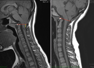
Fig. 1. Cranio-cervical MRI scans from case 18155: preoperative images from 2016 versus postoperative images from 2026. The descent of the cerebellar tonsils and their subsequent ascent are highlighted.
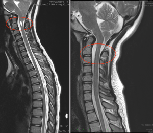
Fig. 2. Cranio-cervical and thoracic MRI scans from case 18155: preoperative images from 2016 versus postoperative images from 2026. The syringomyelic cavity at C2 and its disappearance on the postoperative follow-up scan are highlighted.
Radiological follow-up 9 years after Sectioning of the Filum Terminale
Follow-up MRI scans performed in 2026—nine years after the sectioning of the Filum Terminale according to the Filum System®—show changes compared to the preoperative images.
The main findings are:
- Resolution of the syringomyelia.
- Ascent of the cerebellar tonsils.
- The remaining findings show no significant changes compared to the preoperative images.
Comparison of the MRI scans allows for the observation of the radiological evolution regarding the descent of the cerebellar tonsils and the syringomyelic cavity over the nine-year follow-up period.
Patient’s assessment of the treatment outcome
In her subjective assessment of the results obtained, the young patient reports an 80% improvement in her symptoms and considers the intervention to have been very beneficial.
She also states that she would choose the intervention again—a decision originally made by her parents—and affirms that she recommends the Filum System®.
Favourable long-term evolution
The evolution of this clinical case shows a clinical and radiological improvement maintained during nine years of follow-up after the application of the Filum System® in a paediatric patient.
In the check-up carried out at age 21, a significant decrease in symptoms was observed, along with complete or partial recovery of various objective neurological signs. Changes were also observed in the Magnetic Resonance images, most notably the ascent of the cerebellar tonsils and the disappearance of the syringomyelic cavity described before the intervention.
This case constitutes an example of the long-term evolution observed in a paediatric patient with Arnold-Chiari I Syndrome and idiopathic syringomyelia following the application of the Filum System®, with a favourable subjective assessment of the results and a reported improvement in her quality of life.