
Last update: 03/20/2026, Dr. Miguel B. Royo Salvador, Medical Board number 10389. Neurosurgeon y Neurologist.
Diagnosis
The final part of the spine is composed of 5 vertebrae fused together at the sacral level and by another 3-5 vertebrae fused at the coccygeal level. The sacrococcygeal joint is located between the sacrum and coccyx, each behaving as independent bone structures.
This type of dislocation may occur only when the coccyx is exposed to an intense force, like following a fall or a strong trauma in the gluteus, a birth with significant disproportion between the size of the child and the mother’s pelvis, etc.
The main symptom that a sacrococcygeal dislocation can provoke is pain in the sacrum area, or coccydynia. As the coccyx ligaments receive many nervous fibres, their tearing can produce intense pain. Furthermore, once activated, the pain transmission can trigger an important inflammation in the area.
The diagnosis is usually clinical and based on the typical pain and clinical history, within the circumstances that produce the dislocation.
This type of dislocation is the least diagnosed through complimentary tests. As a matter of fact, the full spine and lumbo-sacral X-rays and MRIs that are performed for other actual or potential diagnostic pictures, do not show the coccyx from S2 vertebra downward. Even in the case of a full visible coccyx, at this level it can be difficult to identify the separation through an X-ray and a CAT or an MRI may be necessary.
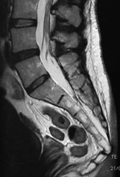
Fig 1. Sacrococcygeal dislocation
Complications
The main risk of a sacrococcygeal dislocation is that it can take a long time to heal and, meanwhile, the pain can limit significantly the patient’s quality of life..
– According to the Filum System®:
At our Institute, in cases of sacrococcygeal dislocation and in some, far less frequent, with fracture of this area, we observe that in most patients it is associated with a diagnosis of Neuro-cranio-vertebral Syndrome (NCVS).This is therefore considered as acquired and traumatic, as it implies an anomalous non-congenital traction of the Filum Terminale. The fracture/luxation of the flexed coccyx, in fact, causes a leverage mechanism, which stretches the filum terminale in its point of insertion located at the first/second coccygeal vertebra level.
The NCVS and its clinical picture can therefore result into a complication of the dislocation, further worsening the patients’ quality of life. The symptoms, in these cases, do not only consist of the typical pain and inflammation in the sacrococcygeal region, but may affect the rest of the spine and the central nervous system, as it occurs in the Filum Disease.
Treatment
– The usual treatment for sacrococcygeal dislocations is conservative: it is recommended to avoid overloading the spine with weight on the coccyx area, to avoid or reduce prolonged sitting, to use pillows or other to reduce the strain while sitting; furthermore indicated are a painkiller and anti-inflammatory therapy, postural physiotherapy or osteopathic manipulation in the coccyx region.
– In the most severe cases, when the pain does not diminish and limits the patient’s usual activity excessively, the treatment can be surgical and it consists of the neurolysis of the affected nerves, or even the removal of the coccyx itself.
According to the Filum System®:
With the luxation or fracture of the sacrum, coccyx or sacrococcygeal, associated to the Neuro-Cranio-Vertebral Syndrome, the Filum terminale ligament that, with others, holds the vertebrae together at this level, is overly tense and exerts an anomalous traction force over the cord and the whole central nervous system. For this reason, after studying these cases for several years, at our Institute we apply a surgical treatment according to the Filum System® method, which includes the sectioning of the Filum Terminale with an exclusive and minimally invasive technique.
The sectioning of the Filum Terminale
References
- M. B. Royo-Salvador, M.V. Fiallos-Rivera, P. Villavicencio (2023), “Neuro-cranio-vertebral syndrome related to coccygeal dislocation: A preliminary study” (PDF).
- Hamoud K, Abbas J (2015) ‘Fracture dislocation of the sacro-coccygeal joint in a 12 year-old boy. A case report and literature review.’, Orthopaedics & Traumatology, Surgery & Research, 101(7), pp. 871-873
- M. B. Royo-Salvador (2014), “Filum System® Bibliography” (PDF).
- M. B. Royo-Salvador (2014), “Filum System® Guía Breve”.
- Stuart RM, Song SJ. (2004) “Unilateral lumbosacral facet joint dislocation without associated fracture.”, Australasian Radiology, 42(2), pp. 224-229.