
Published by ICSEB at 5 May, 2023
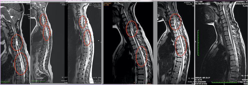
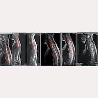
Figure.1- A patient’s magnetic resonances and their evolution following the Filum System® treatment, from the first preoperative MRI until the 7-year postoperative check-up, which shows that the medullary cysts disappeared.
Syringomyelia consists of cystic cavities within the spinal cord usually associated with a generalized neurological deterioration. The most frequent symptoms are pain in the extremities, neck pain, alteration of thermal and tactile sensitivity, lower back pain, upper back pain, headache, gait disorders, paresis, sphincter alterations. Living with this condition means dealing with chronic pain and a progressive loss of autonomy.
Our Institute specializes in the diagnosis and treatment of primary or idiopathic Syringomyelia, which, according to Dr M. B. Royo-Salvador’s caudal traction theory, is caused by the Filum Disease. Hence, the syringomyelic cavity is considered to be a consequence of the ischemia produced by the medullary traction that is generated by a tight filum terminale, non-detectable in the neuroimaging.
The treatment we propose to patients at our centre consists in sectioning the Filum Terminale with a minimally invasive technique, according to the exclusive Filum System® health method. The treatment is applied with the objective of eliminating the root cause of the medullary cavities and stopping the progression of the disease.
After 30 years of interventions since Dr Royo’s discovery of the etiopathogenic mechanism of the disease in 1993, our neurosurgical team continues to see that this condition can be treated with excellent results by applying the Filum System® health method.
“Actually”, says Dr. M. Fiallos, one of the neurosurgery team’s specialists: “not only is the progression of the syringomyelia stopped, preventing the cysts from capturing medullary tissue and its necrosis from expanding – as our patients’ postoperative MRIs show in the short term – but there are also cases in which MR images show how the cavities collapse and gradually disappear in the medium and long term, not appearing new ones (Fig.1). Sometimes, no traces of the disease are left in the neuroimaging and in parallel we can observe the patient’s clinical improvement.”
Thanks to the treatment that we apply at our centre, the correct blood circulation in the central nervous system is restored: “the results that we have obtained so far are not immediately visible, since they are the indirect consequence of a surgical act. However, they are very satisfactory, both objectively for the neurosurgeons and subjectively for the patients. We perform postoperative check-ups 7-10 years after the intervention approximately and can confirm that the cavities shrink and patients clinically recover”, says the neurosurgeon.