
Published by ICSEB at 23 May, 2025
Case No- 18015
Diagnosis: Neuro-Cranio-vertebral Syndrome, Filum Disease. Descent of the Cerebellar Tonsils (Arnold-Chiari Syndrome I). Idiopathic Syringomyelia and idiopathic Scoliosis.
Date of the Sectioning of the Filum terminale (SFT) procedure: April 2015
Patient no. 18015, who wishes to remain anonymous but has given consent for her case to be published, underwent surgery using the Filum System® method at our centre at the age of 7, in 2015.
Before the intervention, her Magnetic Resonance Imaging (MRI) scans, dated 25/03/2015, showed: Filum Disease and Neuro-Cranio-vertebral Syndrome with descent of the cerebellar tonsils (Arnold-Chiari Syndrome I), idiopathic Syringomyelia and Scoliosis. The scoliogram (full spine x-ray) showed: dextroconvex scoliosis at the dorsal level and levoconvex at the lumbar level, and straightening of the cervical lordosis.
In her clinical history, already from the first-year postoperative check-up, a reduction of the intramedullary cavity was recorded. This continued to improve further in the following years, up to the present day, when C. visits ICSEB for her 10-year follow-up.
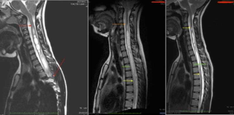
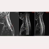
Fig.1 Comparative follow-up of C.: magnetic resonance imaging (25/03/2015 – 15/05/2023 – 19/03/2025)
In her comparative MRI follow-up, the following can be observed:
- In the first preoperative image of Fig.1 (25/03/2015), red arrows are visible. The upper arrow indicates the beginning of the intramedullary cavity at C1, and the lower arrow points to the dorsal scoliosis, which prevents visualisation of the spinal cord.
- In the second image, from her follow-up 8 years after the operation (MRI dated 15/05/2023), there is an improvement in the syringomyelic cavity, along with the disappearance of the dorsal scoliosis.
- In the third image, from her 10-year follow-up after the SFT (MRI dated 19/03/2025), the improvement of the intramedullary cyst continues favourably, with the cavity becoming increasingly filiform.
Clinically, the patient showed improvement from the first postoperative year. Some symptoms improved, others remained unchanged, and others no longer appeared. In her neurological examination, some signs showed recovery, while others persisted without change.
Currently, at 17 years of age and at the 10-year postoperative check-up, C. reports that she no longer experiences the majority (90%) of the preoperative symptoms. The specialist highlights the recovery of most neurological signs, along with the notable reduction of the intramedullary cavity and the disappearance of the scoliotic curvature.
The Filum System® aims to stop the deterioration and progression of Filum Disease and the included diagnoses—as seen in relation to the descent of the cerebellar tonsils, which in this case remains unchanged.
However, practice, most cases achieve better-than-expected results. We observe improvement and relief many symptoms and signs in the majority patients; and in some cases, even reversibility of the visible lesions in the complementary tests, as can be observed in this case.