Last update: 24/03/2023, Dr. Miguel B. Royo Salvador, Medical Board number 10389. Neurosurgeon y Neurologist.
The spine is composed of 33 vertebrae: 7 cervical vertebrae, 12 thoracic or dorsal vertebrae, 5 lumbar vertebrae, 5 sacral vertebrae and 4 or 5 coccygeal vertebrae. The sacral and coccygeal vertebrae form independent bones that are connected through a joint. The other vertebrae, except the first and second cervical vertebrae, are connected through intervertebral discs, interapophyseal joints and ligaments. These joints allow a high mobility and the possibility for humans to stretch, bend and twist.
Vertebral bodies, the most important vertebral structures, support the vertebrae on the front and are separated by intervertebral discs. On the back and on both sides, they are connected through interapophyseal or facet joints, the comfort of which depends on the height of the intervertebral discs.
The structures that form the facet joint are: the fibrous capsule, the synovial membrane, the hyaline cartilage and the bone. Facet joints are innervated by the medial branch of the spinal dorsal ramus of the intervertebral roots, and each of them is dually innervated by the adjacent upper, lower and contralateral segments.
If the height of the intervertebral disc decreases, the friction and limitations it generates between bones excessively degrade articular cartilages. In addition, an extension or separation of the facet joint capsule can occur, which irritates the joints, determining the so-called Facet syndrome or Facet joint syndrome.
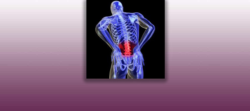
When interapophyseal joints are compressed and worn, the Facet joint syndrome can cause mainly cervical, thoracic or lumbar pain. Depending on the affected facets, pain can be unilateral or bilateral.
Although the pain is mainly located in the lumbar spine, it can also radiate to buttocks, groin and posterior side of thighs, producing lumbago. It can radiate to lower extremities and upper extremities as well. The same can occur in the cervical and thoracic regions, with pain radiating to the upper extremities and thoracic-abdominal region respectively.
The loss of sensitivity in arms and legs is another symptom that can appear. The inflammation of facet joints can also cause stiffness, difficulty in standing up and straight from a sitting position.
On the one hand, pain typically increases with physical exertion or carrying heavy loads, or when maintaining a prolonged standing position, with rotation, bending or contralateral hyperextension. On the other hand, pain can improve when lying down or bending forward.
A facet joint syndrome is suspected when a patient presents cervicobrachialgia, dorsalgia or lombosciatica (even when at rest) and other typical symptoms, with negative neurological and complementary examinations and when no cause is visible on the Magnetic resonance, CT-scan, or Electromiography.
In this case a diagnostic infiltration is performed. This test is carried out when the patient is experiencing her/his usual pain, by administering a local anaesthetic in the proximity of the nerve of the interapophyseal joint that is suspected to be responsible for the pain, with the help of a TV-RX device.
If pain does improve with the infiltration, then it means it is a Facet joint syndrome.
The lumbar facet joint syndrome is mainly caused by the degenerative changes of vertebral joints and joint tissues, as it also happens with osteoarthrosis, cartilage degeneration, etc., which occur over time in interapophyseal joints.
The syndrome may also be due to joint functional alterations, such as the collapse of the intervertebral disc space, with or without disc herniations, blocking and limitation of movement.
Ultimately, it can be caused by other conditions, such as herniations or disc protrusions, cysts, etc.
Lumbar facet joints can be affected by:
The lumbar facet joint syndrome can be complicated mainly by the persistence of its symptoms, which can become chronic if they are not treated, affecting the patient’s quality of life.
Generally speaking, the following treatments can be indicated for the Facet joint syndrome:
– Conservative treatment: several techniques can be applied, including joint repositioning through the mobilization of the spine and physical therapy exercises aimed at muscles’ flexibility and strength. If the physical therapy is not sufficient, anti-inflammatory medication or cortisone is administered.
– If the conservative treatment fails, only in selected cases the treatment can focus on the infiltration of anaesthetics and cortisone at the level of the affected joints.
– Definitive pain suppression in the Facet joint syndrome is obtained by producing an injury into the interapophyseal joint nerve, thus eliminating pain transmission. There are various techniques, such as percutaneous neurotomy, which destroys the sensory nerve fibres of the facet joint through radiofrequency, with cauterization of the neurofilaments.
– According to the Filum System®:
The Facet joint syndrome is frequently found in patients diagnosed with Filum Disease. The caudal traction force that generates this condition also affects the vertebral interapophyseal joints, by overloading them and supporting the appearance of arthritic processes. For this reason, before addressing a Facet joint syndrome, we carry out the Filum System® diagnostic protocol to identify a possible Filum Disease, which would need to be treated prior to the Facet joint syndrome.
At out centre, the treatment of choice is electrocoagulation with high frequency current, a technique that we apply for its precision and safety, with excellent results.
Monday to Thursday: 9-18h (UTC +1)
Friday: 9-15h (UTC +1)
Saturday and Sunday closed
Toll-free from the US: +18882012544
+34 932 800 836
+34 932 066 406
Legal regulations
Legal notice
Pº Manuel Girona, nº 32
Barcelona, España, CP 08034

The Institut Chiari & Siringomielia & Escoliosis de Barcelona (ICSEB) complies with the established in EU regulation 2016/679 (GDPR).
The contents of this website are a non-official translation of the original content of the website in Spanish. The translation is courtesy of the Institut Chiari & Siringomielia & Escoliosis de Barcelona with the purpose of facilitating comprehension for anyone who wishes to Access the website.