


Last update: 10/21/2026, Dr. Miguel B. Royo Salvador, Medical License Number 10389. Neurosurgeon and Neurologist.
Definition
Cerebrospinal fluid (CSF) is a clear, colourless fluid that circulates around the brain and spinal cord, protecting the central nervous system (CNS). It is found in the cerebral ventricles, cavities located within the brain, and also in the subarachnoid space that surrounds the brain and spinal cord.
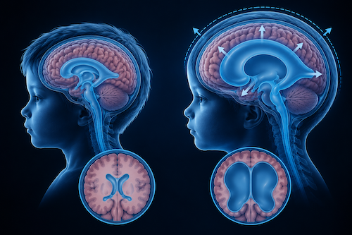 Fig. 1 Left image: brain with normal CSF circulation. Right image: brain with hydrocephalus.
CSF performs important functions, such as protecting the brain and spinal cord, cushioning impacts, and contributing to the balance of the environment in which the cells of the nervous system function. Under normal conditions, it is continuously produced and reabsorbed, maintaining a relatively stable volume and pressure (Fig. 1, left image).
Hydrocephalus is a neurological condition that occurs when there is an alteration in the circulation, production, or reabsorption of CSF, which can lead to its excessive accumulation within the brain ventricles. As a result, the ventricles may enlarge, and in some cases, intracranial pressure may increase (Fig. 1, image on the right).
Hydrocephalus can occur at any age and may arise from various causes, presenting in different types and with diverse clinical manifestations. Consequently, its diagnosis and treatment depend on the individual’s age, the underlying cause of the CSF accumulation, and the specific characteristics of each case.
Symptoms
Symptoms of hydrocephalus can appear acutely or develop progressively. Clinical manifestations vary depending on the individual’s age, the cause of the hydrocephalus, and the rate at which cerebrospinal fluid accumulates.
During pregnancy:
In some cases, hydrocephalus can be detected before birth via prenatal ultrasound, which may reveal enlarged cerebral ventricles in the fetus.
In infants and young children:
One of the most characteristic signs is an increase in head circumference that occurs more rapidly than expected for the child’s age, as the skull bones have not yet fully fused. Other symptoms may include irritability, drowsiness, vomiting, feeding difficulties, or the so-called “setting-sun sign” in which the eyes appear to be directed downward.
In children:
Symptoms of hydrocephalus may include headaches, nausea, vomiting, and visual disturbances. Depending on the cause and progression of the condition, there may also be behavioural changes, drowsiness, or difficulties with school performance.
In adolescents and young adults:
Symptoms may include persistent headaches, visual disturbances, dizziness, problems with balance and coordination, difficulty walking, nausea, episodes of loss of consciousness (syncope), and impairments in memory or other cognitive functions.
In adults and older individuals:
Symptoms may primarily affect gait and balance, cognitive functions, and bladder control. Possible manifestations include difficulty walking or standing, slowed movements, memory and concentration issues, a decrease in interest or initiative, and altered urinary control.
It is important to note that not everyone with hydrocephalus experiences the same symptoms. The intensity and progression of these manifestations depend on several factors, including the age of onset, the underlying cause of the hydrocephalus, and the speed at which it develops. In the presence of consistent symptoms, a medical evaluation is necessary to determine their potential origin.
Furthermore, Hochstetler et al. (1) describe, in general, a clinical picture characterized by “disorientation and pain (headaches), cognitive and developmental changes, vision and sleep disturbances, and gait abnormalities.”
Diagnosis
Kahle et al. note that “Beyond infancy, hydrocephalus typically presents with a constellation of findings that include some combination of headache, vomiting, loss of developmental milestones, diplopia (usually from a VI cranial nerve palsy), or papilloedema.”
Following a neurological examination with these findings, a specialist may order “brain imaging—the most important diagnostic tool” (2)—which includes:
– ultrasonography,
– computed tomography (CT),
– magnetic resonance imaging (MRI).
Magnetic resonance imaging (MRI) is generally indicated to clarify the anatomy and determine the cause of the hydrocephalus. It is preferred over computed tomography (CT) because MRI avoids radiation exposure and provides a greater amount of information.
Cine-MRI (or a cine-MRI study of CSF flow) can provide information regarding patient-specific changes in cerebrospinal fluid dynamics.” (2)
In adults, additional procedures may include lumbar puncture, continuous lumbar CSF drainage, intracranial pressure monitoring, a lumbar infusion test (to measure CSF outflow resistance), or radionuclide cisternography.
Types
Hydrocephalus can be classified based on the circulation of cerebrospinal fluid (CSF), its underlying cause, and the time of onset.
- Communicating hydrocephalus: CSF circulates through the ventricles, but there is an impairment in its reabsorption.
- Obstructive or non-communicating hydrocephalus: an obstruction hinders the normal circulation of CSF
- Primary normal-pressure hydrocephalus: typically occurs in older adults with no identifiable cause.
- Secondary normal-pressure hydrocephalus: develops as a result of a known cause, such as a hemorrhage, trauma, or infection.
- Congenital or primary hydrocephalus: present at birth or develops during the fetal period.
- Acquired or secondary hydrocephalus: develops after birth as a result of another medical condition, injury, or other cause.
“The normal flow of CSF is from lateral ventricles through the foramen of Monroe, into the common third ventricle proceeding into the fourth ventricle through the cerebral aqueduct, and out of the foramen of Luschka and Magendie into the subarachnoid space.
Communicating hydrocephalus occurs when bulk flow is unobstructed and results from a failure to absorb CSF via the normal drainage pathways or an accumulation of CSF due to an over-production.
As the name suggests, non-communicating hydrocephalus occurs when there is an obstruction to the normal CSF flow through the central nervous system (CNS), and this can be due to a number of precipitating causes.
Despite seemingly simple diagnostic criteria such as ‘communicating’ versus ‘non-communicating’—among other classifications— the reality is that there remains a burden of clinical heterogeneity among patients with hydrocephalus.
Attempts to describe and classify hydrocephalus into discrete categories have often resulted in confusion as there is notable overlap when considering popular categories such as ‘congenital’ or ‘acquired’.” (1)
For this reason, Hochstetler et al. prefer to categorize hydrocephalus as either “primary (syndromic and/or idiopathic) or secondary to another disease or condition.” (1)
- Primary hydrocephalus may arise from various genetic factors during fetal development.
- Secondary hydrocephalus can result from numerous causes, including—but not limited to—infections, hemorrhages, and trauma..
Cause
“Hydrocephalus is a serious condition that affects patients of all ages, resulting from a multitude of causes. While the aetiologies of hydrocephalus are numerous.” (1)
The primary causes of hydrocephalus include:
- Congenital hydrocephalus: this can be related to developmental abnormalities of the nervous system during pregnancy, including certain genetic anomalies or issues with fetal development.
- Acquired hydrocephalus: this can develop as a result of traumatic brain injuries, tumors, meningitis or other infections, cerebral hemorrhages, or other conditions that disrupt the circulation or reabsorption of cerebrospinal fluid.
In the case of normal-pressure hydrocephalus, there is an accumulation of CSF associated with an enlargement of the brain ventricles, without necessarily involving a sustained increase in intracranial pressure
- Primary normal-pressure hydrocephalus: in most cases no specific cause for the CSF accumulation ca be identified.
- Secondary normal-pressure hydrocephalus: it can occur a s a result of a known cause, such as brain surgery, a cyst, an injury, a hemorrhage, or an infection.
Risk factors
It is recognized that “contributory factors such as dysfunction of the glymphatic system, diminished arterial pulsatility, metabolic and osmotic dysregulation, astrogliosis and neuroinflammatory processes are acknowledged as critical in the pathogenesis of NPH.” (2)
Complications
This is a complex condition, in which long-term effects vary significantly from case to case.
Various complications may arise, such as limitations in executive functions, cognitive difficulties—particularly regarding short-term memory—vision problems, and chronic headaches. (3)
Secondary dementia in adults is one of the few complications that is treatable and potentially reversible.
If not properly treated, hydrocephalus can be a fatal condition.
Treatment
– Treatment options for hydrocephalus are surgical and are aimed at relieving intracranial pressure:
- Shunt systems: intracranial valves or shunts
These involve the placement of a catheter that directs excess cerebrospinal fluid (CSF) from the lateral ventricles to the peritoneum (ventriculoperitoneal shunt, VPS) or to the right atrium (ventriculoatrial shunt, VAS), where it is absorbed. This bypasses the site of a mechanical obstruction or a functional impairment in CSF flow or absorption. (4)
- Endoscopic third ventriculostomy (ETV)
This procedure involves creating an opening in the floor of the third ventricle to allow CSF to flow into the prepontine cistern. (4)
Hochstetler et al. state that “the diagnosis and treatment of the various forms of adult hydrocephalus remain understudied and underreported. Surgical interventions to treat hydrocephalus, though lifesaving, have a high incidence of failure.
– Previously tested pharmacotherapies for the treatment of hydrocephalus have resulted in net zero or negative outcomes for patients potentially due to the lack of understanding of the cellular and molecular mechanisms that contribute to the development of hydrocephalus.” (1)
According to the Filum System®:
When hydrocephalus occurs in association with Neuro-Cranio-Vertebral Syndrome—or Filum Disease—which encompasses Arnold-Chiari syndrome type I and/or idiopathic syringomyelia, the Filum Terminale ligament is found to be excessively tense. This exerts an abnormal traction force on the spinal cord and, consequently, on the entire central nervous system. This force leads to the herniation of the cerebellar tonsils through the foramen magnum (occipital opening), the development of tentorial herniations, and the narrowing (stenosis) of the cerebral aqueduct (aqueduct of Sylvius) due to this traction.
For this reason, based on years of clinical observation—including in cases involving associated hydrocephalus—our Institut Chiari de Barcelona employs a surgical approach according to the Filum System®. This method involves the sectioning of the Filum Terminale using a proprietary and exclusive, minimally invasive technique. This procedure releases the central nervous system from the traction force, alleviating the stenosis and improving the circulation of cerebrospinal fluid.
Therefore, in cases presenting with hydrocephalus, Arnold-Chiari syndrome type I, and/or idiopathic syringomyelia, we first proceed with the sectioning of the Filum Terminale. This is typically sufficient to improve the hydrocephalus, often precluding the need for further treatment.
Results
The results of surgical treatment for hydrocephalus are generally very positive; in most cases, they allow for the restoration of a normal life expectancy and the recovery of lost functions. (3)
Each of the aforementioned treatments also carries potential complications:
– Shunts (valves) can become obstructed or fail, which may lead to a critical clinical situation if immediate action is not taken. The primary complications associated with CSF shunts include infections, insufficient drainage due to mechanical failure, and overdrainage.
– Endoscopic third ventriculostomy by contrast, has a success rate ranging from 30% to 70%. Due to the high failure rate of this procedure, attempts have been made to combine it with the simultaneous cauterization of the choroid plexuses. (4)
-In children, subcutaneous tunneling is associated with fewer complications compared to subcutaneous suturing. Both surgical experience and the operative setting significantly influence outcome, highlighting the importance of careful technique selection and supervision in pediatric external ventricular drain placement” (5)
Application of the Sectioning of the Filum terminale according to the Filum System®:
Case Nº 17076
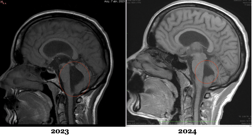
Fig.2. Cranial magnetic resonance imaging (MRI) scans of an ICSEB patient, before and after the sectioning of the filum terminale according to the Filum System®:
- 2023: preoperative MRI showing hydrocephalus.
- 2024: postoperative MRI showing improvement in the hydrocephalus.
References
- Hochstetler, A., Raskin, J., & Blazer-Yost, B. L. (2022). Hydrocephalus: Historical analysis and considerations for treatment. European Journal of Medical Research, 27, 168. https://doi.org/10.1186/s40001-022-00798-6
- Kahle, K. T., Kulkarni, A. V., Limbrick, D. D., Jr., & Warf, B. C. (2016). Hydrocephalus in children. The Lancet, 387(10020), 788–799. https://doi.org/10.1016/S0140-6736(15)60694-8
- Hydrocephalus Association. (s. f.). La hidrocefalia. https://www.hydroassoc.org/la-hidrocefalia/
- Moreno Mendieta, M., & Núñez Enamorado, N. (2022). Hidrocefalia en pediatría. Protocolos Diagnóstico Terapéuticos de la AEP: Neurología Pediátrica, 1, 145–150.
- Araceli, T., Klarner, S., Zeman, F., Haj, A., Siller, S., Proescholdt, M., Schmidt, N. O., & Ott, C. (2026). Complication profile of two surgical techniques, neurosurgical experience, and operative setting for pediatric external ventricular drainage implantation. Child’s Nervous System, 42, 330. https://doi.org/10.1007/s00381-026-07413-7
- Bubenikova, A., Procházka, V., Vacínek, D., et al. (2026). [Título del artículo]. Journal of Neurology, Neurosurgery & Psychiatry. https://doi.org/10.1136/jnnp-2025-336076