
Published by ICSEB at 4 July, 2025
Case 19475
Diagnosis: Neuro-cranio-vertebral Syndrome. Filum Disease. Descent of the cerebellar tonsils (Arnold-Chiari I Syndrome). Intramedullary cyst (Idiopathic Syringomyelia).
Date of the Sectioning of the Filum Terminale (SFT) intervention according to the Filum System®: April 2018.
The patient, who wishes to remain anonymous, was operated on at the age of 11 and has since returned to our centre for several follow-ups, the last one being 7 years after the intervention, in early 2025.
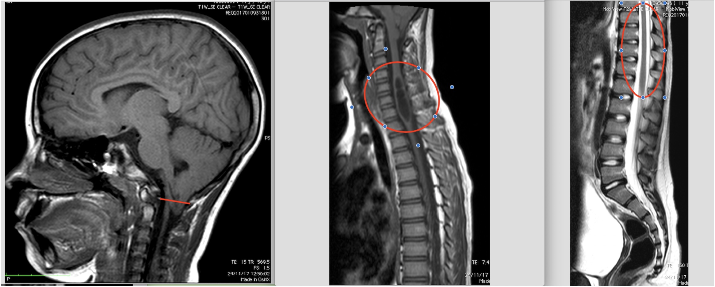
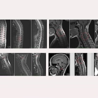
Fig.1. Preoperative images of Case 19475: magnetic resonance scans from the end of 2017 – brain, cervical, thoracic and lumbo-sacral.
Before the application of the treatment, clinically she suffered especially from headaches, instability and vertigo, easy and generalised fatigue.
In her preoperative MR images from 2017 (Fig.1), the following findings, among others, could be observed:
- Descent of the cerebellar tonsils between the lower edge of the posterior arch of C1 and the upper edge of the posterior arch of C2. Mild dilation of the fourth ventricle.
- Syringomyelic cavity at C4-D1, highly dilated.
- Cervical and thoracic ischaemia-oedema, extending to the conus medullaris (pre-syringomyelia).
Since the check-up one month after surgery, and the one-year follow-up (2019), and then all subsequent ones (2023 and 2024), the patient showed an overall improvement in all symptoms, up to the near-complete disappearance of symptoms at the last visit (2025). Similarly, with regard to the neurological examination, most of the objective clinical signs had recovered.
In this case, we can observe how also in the magnetic resonance imaging, the images correspond to the progressive postoperative improvement in the patient’s condition, as seen in Figures 2, 3, and 4.
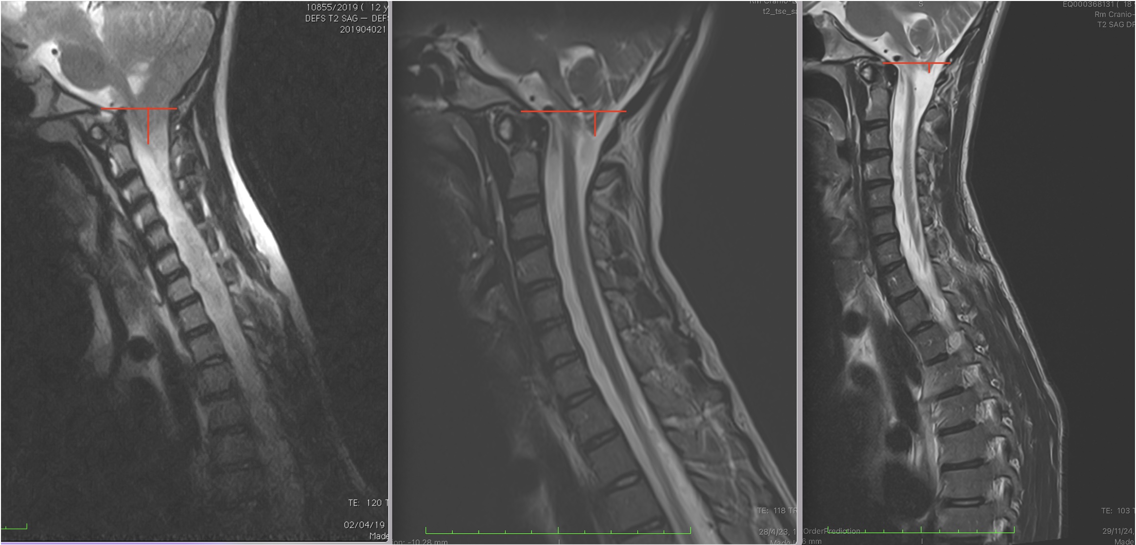
Fig.2 Comparative follow-up of Case 19475: control MRIs from 2019-2023-2024: ascent of the cerebellar tonsils.
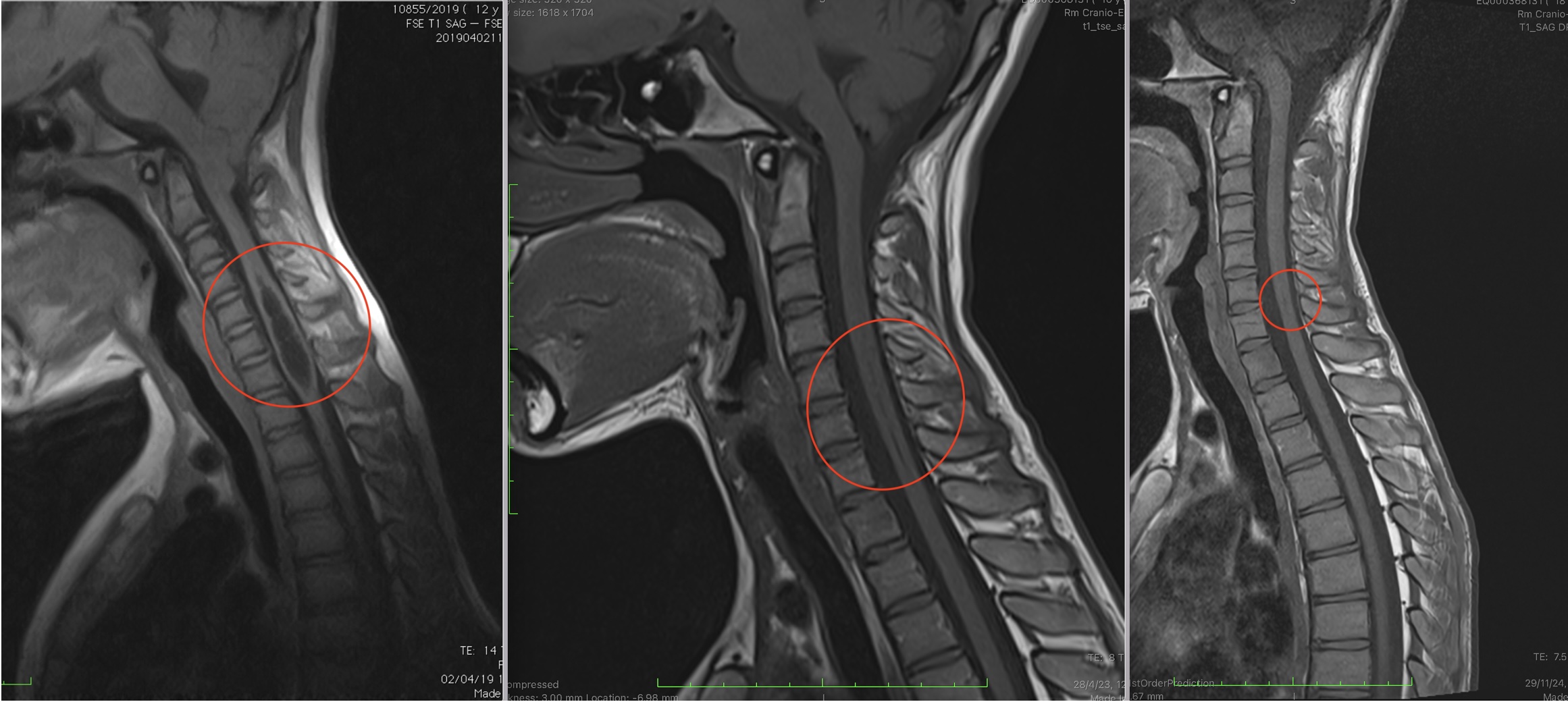
Fig.3 Comparative follow-up of Case 19475: control MRIs from 2019-2023-2024: reduction of the cervical syringomyelic cavity.
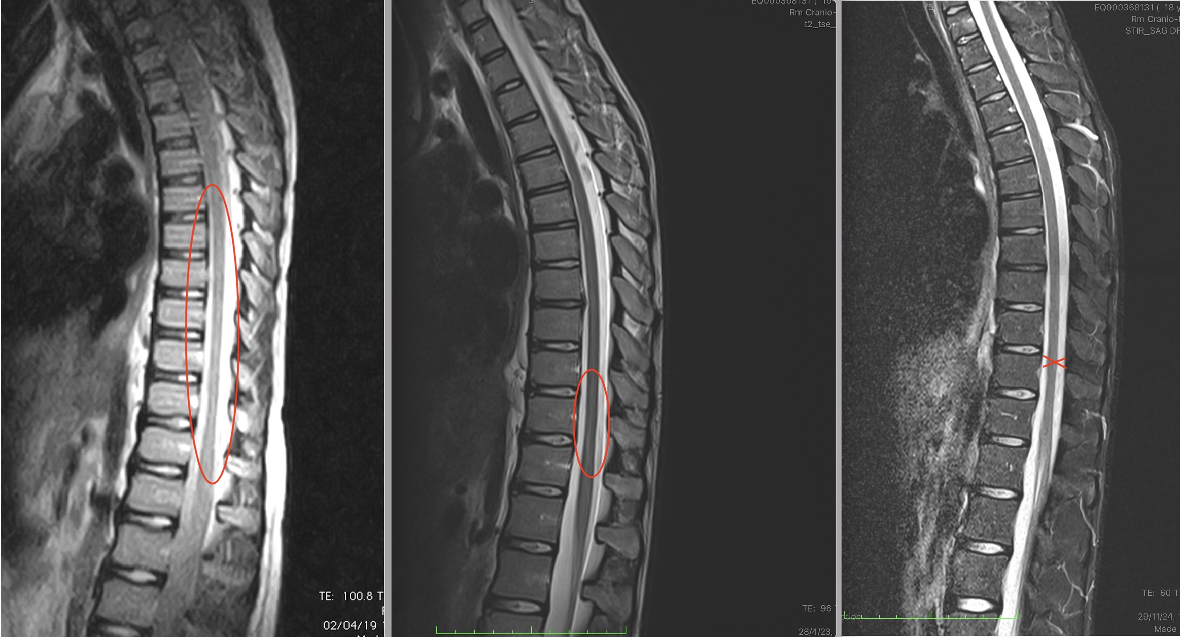
Fig.4 Comparative follow-up of Case 19475: control MRIs from 2019-2023-2024: reduction of the thoracic and lumbar ischaemia/oedema (pre-syringomyelic image).
Here we present, as the first image in each follow-up comparison, the MRIs from 2019, which were still unchanged compared to the preoperative ones from 2017. In most cases, one must wait a few years before visible improvements appear in the images.
Starting from the control MRIs of 2023, five years after the intervention, we have clear evidence of visible improvement in each of the images, even greater in the most recent follow-up of the end of 2024, seven years post-op, showing:
- Ascent of the cerebellar tonsils and normalisation of the fourth ventricle (Fig.2).
- Syringomyelia from C5 to C7, with a reduction also in diameter (Fig.3).
- Reduction of the ischaemia/oedema image in the thoracic region (Fig.4).
We are delighted with the excellent outcome of the treatment in this patient’s case, who now, at the age of 18, is able to enjoy the opportunity that releasing the spinal cord from the traction caused by the Filum terminale has given her. We wish her continued well-being.
The Filum System® halts the progression of Arnold-Chiari I Syndrome and Idiopathic Syringomyelia, promoting the restoration of reversible lesions in the nervous system and improving patients’ quality of life.