El comentario del Dr. Zuev, neurocirujano ruso, en un forum:
(se puede encontrar el comentario original en ruso en el link: https://www.syringomyelia.ru/forum/–/160—–?limit=10&start=10)
Traducción del comentario:
Вuenos días a todos quien se interesa por ese tema!
Finalmente hemos podido encontrar unas publicaciones internacionales de los institutos médicos de “referencia” en America y Europa, que desmienten teorías del origen y patogenia de SACH y SM, de los que está “gritando” el Institut en Barcelona. Noten que los artículos han sido publicados en las revistas respetables:
1. “To date, there are no published reports of an explicit relationship between CM-I and TCS. In a recent study of spinal cord traction in fresh cadavers, caudal tension on the CMD was found to produce less than 1 mm downward movement of the medulla and upper cervical spinal cord and no displacement of the cerebellar tonsils. The authors concluded that caudal fixation of the spinal cord is an implausible cause of CM-I and that SFT is unlikely to reverse tonsillar ectopia” – Tubbs RS, Loukas M, Shoja MM, Oakes WJ. Observations at the craniocervical junction with simultaneous caudal traction of the spinal cord. Childs Nerv Syst 2007;23:367-9.
2. This cord traction theory was generally dismissed as an oversimplification
that did not account for the complex neural as osseus anomalies of Chiari malformation type II and did not explain why nerve roots below the mid-thoracic level coursed normally rather than cephalad as would be expected
if there was significant downward traction on the CMD – Association of Chiari malformation type I and tethered cord syndrome: preliminary results of sectioning filum terminale. – Thomas H. Milhorat et al.- Surgical Neurology 72 (2009) 20–35 – Department of Neurosurgery, The Chiari Institute, Harvey Cushing Institutes of Neuroscience, USA
3. In patients with large tonsillar herniations extending below the arch of C1, it is our practice to perform a posterior fossa decompression before SFT to avoid the potential risk of exacerbating tonsillar herniation by the
sudden release of lumbar CSF. – Association of Chiari malformation type I and tethered cord syndrome: preliminary results of sectioning filum terminale. – Thomas H. Milhorat et al.- Surgical Neurology 72 (2009) 20–35 – Department of Neurosurgery, The Chiari Institute, Harvey Cushing Institutes of Neuroscience, USA
En el último artículo hay mucho de interés sobre el tema pero el resumen es el siguiente: LA SECCIÓN DEL FILUM TERMINALE DEBE SER REALIZADA SÓLO EN EL CASO RIGUROSAMENTE INDICADO y no a todos los enfermos con SACH y SM.
La respuesta del Dr. Royo Salvador al comentario:
Sr. Zuev hay que puntualizar en su comunicación algunas consideraciones en relación a la publicación de Tubbs, como:
1. “Tubbs intenta con una experimentación inadecuada replicar a mi artículo del 2005”
El artículo que cita: “Tubbs RS, Loukas M, Shoja MM, Oakes WJ. Observations at the craniocervical junction with simultaneous caudal traction of the spinal cord. Childs Nerv Syst 2007;23:367-9.”
Quiere ser una réplica a otro publicado: “Royo-Salvador MB, Solé-Llenas J, Doménech JM, Gonzalez-Adrio R (2005). Results of the section of the filum terminale in 20 patients with syringomyelia scoliosis and Chiari malformation. Acta Neurochir (Wien) 147:515–523.”
Vease la bibiliografía del artículo de Tubbs:
1. Goldstein F, Kepes JJ (1966) The role of traction in the development of the Arnold-Chiari malformation. An experimental study. J Neuropathol Exp Neurol 25:654–666
2. Oakes WJ, Tubbs RS (2004) Chiari malformations. In: Winn HR (ed) Youmans neurological surgery, 5th edn. Saunders, Philadel- phia, pp 3347–3361
3. Royo-Salvador MB, Solé-Llenas J, Doménech JM, Gonzáalez- Adrio R (2005) Results of the section of the filum terminale in 20 patients with syringomyelia scoliosis and Chiari malformation. Acta Neurochir (Wien) 147:515–523
4. Tubbs RS, Salter G, Grabb PA, Oakes WJ (2001) The denticulate ligament: anatomy and functional significance. J Neurosurg 94 (Spine 2):271–275
5. Tubbs RS, Bui CJ, Rice WC, Loukas M, Naftel RP, Holcombe MP, Oakes WJ (2006) Critical analysis of the Chiari I malformation found in children with lipomyelomeningocele. J Neurosurg (in press).
En ese artículo de Tubbs para concluir y dar la respuesta a mi artículo, cita:
“Royo-Salvador et al. [3] have described three patients with CIM, two of these with syringomyelia, in whom symptoms were improved in two after transection of the filum terminale. Interestingly, these patients did not have a fatty infiltrated filum terminale, low conus medullaris, and were operated “without opening the dural sac.”
Los autores no se han tomado la molestia de leerse el fundamento teórico previo a ese artículo, que es mi tesis doctoral “Aportación a la etiología de la Siringomielia” de 1992, y tres publicaciones más:
Royo-Salvador MB. [A new surgical treatment for syringomyelia, scoliosis, Arnold-Chiari malformation, kinking of the brainstem, odontoid recess, idiopathic basilar impression and platybasia]. Rev Neurol. 1997 Apr;25(140):523-30. Spanish.
Royo-Salvador MB. [Platybasia, basilar groove, odontoid process and kinking of the brainstem: a common etiology with idiopathic syringomyelia, scoliosis and Chiari malformations]. Rev Neurol. 1996 Oct;24(134):1241-50. Spanish.
Royo-Salvador MB. [Syringomyelia, scoliosis and idiopathic Arnold-Chiari malformations: a common etiology]. Rev Neurol. 1996 Aug;24(132):937-59. Review. Spanish.
Posiblemente porque los autores solo leen en norteamericano y se han olvidado o desconocen de que su Medicina se la enseñamos nosotros, puesto que es un producto de la cultura mediterránea.
En 1975 cuando iniciamos la investigación de la tracción del sistema nervioso como causa de la siringomielia idiopática y el SACH.I con el Dr. Domenech catedrático de la cátedra de Anatomía y Embriología Humana de la Facultad de Medicina de la Universidad Autónoma de Barcelona, descartamos el estudio con cadáveres, puesto que su comportamiento es diferente al “vivo”: la elasticidad, las compensaciones del conjunto neuro-cráneo-vertebral, la inflamación, la circulación de los líquidos intersticiales, del LCR… De igual forma descartamos la experimentación animal, ya que siempre se podría decir que los hallazgos no son extrapolables al humano por cualquier mínima razón. De hecho iniciamos una simulación de la tracción medular con embriones de pollo que no resultó viable.
Al final me decidí por la observación en los pacientes y demostrar la existencia de una fuerza de tracción medular en pacientes afectos de Siringomielia idiopática, al demostrar estadísticamente que la posición de su cono medular era más baja que en la población normal, mediante estudios de RM de toda la columna vertebral en 292 pacientes y 50 sin las enfermedades estudiadas (“Aportación a la etiología de la Siringomielia”, 1992, Universidad Autónoma de Barcelona, España).
En el artículo de Tubbs, que lo conocemos desde la fecha de su publicación, se aplica una fuerza de 75N/16 lbs y la observación para la deformación es “simultánea” en la región cervical, en tronco cerebral y en las amígdalas cerebelosas: “In 12 fresh adult cadavers (less than 6 h postmortem), distal tension (75 N/16 lbs) utilizing a manual tension gauge (Lyman, Middletown, CT, USA) was applied to the conus medullaris with simultaneous observation of the cervical spinal cord, brainstem, and hindbrain (cerebellar tonsils) and their relationship to the foramen magnum (referenced to a metric ruler placed at the foramen magnum after the removal of the posterior arch of the atlas and 2 cm of the midline occiput”.
Para valorar una parte inadecuada de ésta experimentación cuya conclusión se quiere aplicar a la clínica humana, hay que considerar la fórmula de la elongación de un cuerpo físico, que es:
X = A.cosw.t
(donde X es la elongación, A el radio, w=2pi/T (T=periodo) y t=tiempo).
Para el cálculo de la elongación un factor directamente proporcional es “t”, el tiempo de aplicación de la fuerza. Si consideramos que un año tiene 31.536.000 segundos y muchos de los pacientes soportan de forma continuada la fuerza de tracción sin ningún descanso, más de 20-30 años. Así para obtener la elongación durante ese período tendríamos que multiplicar lo observado en un segundo, como la observación simultánea de la publicación de Tubbs, por 946.080.000 que son los segundos que tienen 30 años.
En palabras más sencillas, la elongación del sistema nervioso por una fuerza aplicada a la médula espinal depende también del tiempo que se aplica; no es lo mismo la elongación resultado de aplicar una fuerza durante 1 segundo ó 5 minutos ó durante 30 años.
Le sorprende a Tubbs que en mi publicación no se realice apertura del saco dural para la sección del filum terminale, “and were operated “without opening the dural sac””, eso indica que desconoce datos anatómicos importantes. En una determinada zona del filum terminale externo es posible seccionar la inserción conjunta del filum terminale ó también llamado ligamento coccígeo, y la inserción de la duramadre.
En cuanto a la cita de Milhorat:
En el año 2004 tuve una reunión personal y profesional con el Dr. Thomas H. Milhorat, en un hospital de Nueva York, donde intercambiamos información, opiniones y teorías en relación al SACH.I, Siringomielia y Escoliosis idiopática. Es significativo que el hasta entonces paladín de la craniectomía suboccipital aplicada al SACH.I por la teoría de la fosa craneal pequeña, cambiara su discurso he iniciara la aplicación de la SFT en el SACH.I. De tal forma que el siguiente artículo que publicó tras nuestro encuentro es el que se cita:
“Association of Chiari malformation type I and tethered cord syndrome: preliminary results of sectioning filum terminale. – Thomas H. Milhorat et al.- Surgical Neurology 72 (2009) 20–35”.
Este artículo desde hace algunos años figura comentado en nuestra web.
Hay varios hechos significativos:
Milhorat copió parcialmente mi teoría sin realizar ningún estudio científico previo que justificara su cambio de criterio y actuación, tampoco dijo que la teoría en la que se basaba ahora era de otro, la hizo suya como hacen frecuentemente algunas personas.
Al aplicar la SFT en pacientes con el SACH.I, Milhorat comprueba que es útil en la siringomielia. Pero ignora por completo la aplicación para la Escoliosis idiopática.
Milhorat no se molestó en leer mis anteriores publicaciones, ni mi tesis, donde se demuestra lo que más tarde aplico en los artículos posteriores de la casuística operatoria. Posiblemente porque los norteamericanos sólo leen en norteamericano. Y no se han enterado que actualmente el idioma español ha superado en uso al inglés.
Milhorat no pudo copiar mi técnica quirúrgica mínimamente invasiva para la SFT, puesto que no se la expliqué, y realizó laminectomías lumbares para la SFT con las consiguientes fístulas de LCR, que le ha causado múltiples demandas judiciales a él y a su equipo incluido Bolognese.
Actualmente Milhorat está jubilado y continua su consulta Bolognese, que no ha publicado ni una página como primer autor en relación al SACH.I, Siringomielia y Escoliosis idiopática. Sus conocimientos y teorías es como la de todos los neurocirujanos que no han realizado investigaciones sobre el SACH.I, la Siringomielia y Escoliosis idiopática. Es decir, Bolognese, está donde estábamos nosotros hace 37 años.
Le recuerdo que nosotros tenemos como primer autor:
“Aportación a la etiología de la siringomielia”. Miguel B. Royo Salvador. 1992. Universidad Autónoma de Barcelona.Tesis Doctoral. Con la estadística más importante del mundo estudiada por un sólo autor, de 292 casos de siringomielias asociadas o no a Chiari I y escoliosis. En forma de 319+55 apéndice=374 páginas de la Tesis doctoral.
Y las publicaciones posteriores en relación al tema: Que suman 58 páginas en tres idiomas: español, francés e inglés. Suman un total de 423 páginas publicadas. La tesis doctoral aprobada “cum laude” por un tribunal de tesis doctoral de seis catedráticos o especialistas significativos. Y el resto de publicaciones por tribunales de la especialidad médicos y éticos de los comités de las diferentes publicaciones.
1: Royo-Salvador MB, Sole-Llenas J, Domenech JM, Gonzalez-Adrio R. Results of the section of the filum terminale in 20 patients with syringomyelia, scoliosis and Chiari malformation. Acta Neurochir (Wien). 2005 May;147(5):515-23; discussion 523.
2: Royo-Salvador MB. [On the relationship between Chiari malformations and hydrosyringomyelia]. Rev Neurol. 1999 Aug 16-31;29(4):389-90. Spanish.
3: Royo Salvador MB. Referring to the posterior fossa cranioectomy and tonsillar resection in order to treat Chiari I malformation with syringomyelia. Acta Neurochir (Wien). 1999;141(9):1020-1.
4: Royo-Salvador MB. [The etiology of the Chiari I/syringomyelia complex]. Neurologia. 1999 Oct;14(8):415-8. Spanish.
5: Royo Salvador MB. [Tonsillectomy as a treatment of Chiari I malformation with syringomyelia]. Neurochirurgie. 1999 Nov;45(4):338-9. French.
6: Royo-Salvador MB. Relating to classification and etiology of Chiari I malformation. Magn Reson Imaging. 1999 Nov;17(9):1403.
7: Royo Salvador MB. “Pseudo” idiopathic scoliosis in syringomyelia. Eur Spine J. 1999;8(5):421.
8: Royo-Salvador MB. [Etiology and treatment of Chiari I/syringomyelia complex]. Rev Neurol. 1999 Jun 16-30;28(12):1218. Spanish.
9: Royo-Salvador MB. [A new surgical treatment for syringomyelia, scoliosis, Arnold-Chiari malformation, kinking of the brainstem, odontoid recess, idiopathic basilar impression and platybasia]. Rev Neurol. 1997 Apr;25(140):523-30. Spanish.
10: Royo-Salvador MB. [Platybasia, basilar groove, odontoid process and kinking of the brainstem: a common etiology with idiopathic syringomyelia, scoliosis and Chiari malformations]. Rev Neurol. 1996 Oct;24(134):1241-50. Spanish.
11: Royo-Salvador MB. [Syringomyelia, scoliosis and idiopathic Arnold-Chiari malformations: a common etiology]. Rev Neurol. 1996 Aug;24(132):937-59. Review. Spanish.
Las múltiples interpretaciones y omisiones sin estudios científicos en relación a nuestras investigaciones, se pueden resumir en:
Que el tipo de publicación anglosajona de un número escaso de páginas por artículo da la idea errónea que es suficiente para entender y aplicar las conclusiones en forma de tratamiento de una teoría compleja como la que exponemos y contraria a la que está en uso.
Cuando la publicación es tan extensa como nuestras dos primeras publicaciones de 22, 9 páginas, y además en español, muchos profesionales no llegan a leer todo el artículo e incluso si lo leen todo, se hacen una idea vaga y propia de lo que se pretende explicar.
En ambos casos anteriores la documentación parcial adquirida, supone para algunos profesionales suficiente como para hacer una teoría propia, aplicando a su manera las conclusiones e interviniendo a los pacientes sin criterios científicos previamente demostrados, con los consiguientes resultados erráticos, que acabará en el desprestigio de una teoría correcta y de la solución.
Por todo lo anterior, unido a la existencia de varios neurocirujanos que aprovechándose de nuestra repercusión mediática dicen hacer lo mismo que nosotros, (por ejemplo ver la imagen de abajo, que no ha tenido ningún contacto con nosotros), hemos considerado:
La creación de la marca registrada Filum System®, donde se recoge lo fundamental de nuestras investigaciones, que se impartirá en la Filum Academy Barcelona®, dando la acreditación a los profesionales que realicen el curso de formación. Desistiendo por ahora en proceder a más publicaciones o participación en Congresos, excepto las que se realicen para el Filum System®.
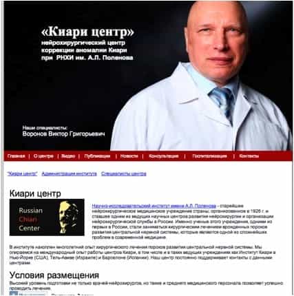
Traducción de la imagen:
“Nosotros nos respaldamos con la experiencia de trabajo de muchos años de los centros Chiari, incluidos los centros líderes como Institut Chiari en Nueva-York (E.E.U.U.), Tel-Aviv (Israel) y Barcelona (España). Nuestro centro mantiene constantemente el contacto con estos centros”.