Maria Maddalena Crabolu. Cord Traction Syndrome. Descent of the Cerebellar Tonsils. Idiopathic Syringomyelia.
Published by ICSEB at 7 March, 2018
Surgery date: 03/12/2013
![]()
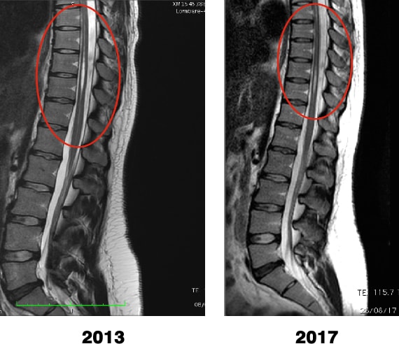
The patient had a magentic resonance imaging check up of the spine for her 5 year post-op follow up of the Sectioning of the Filum Terminale according to the Filum System® method.
Here we can see the comparison betwenn the pre- and current post-op MRI scans with the relevant decrease of the syringomyelic cavity.
Syringomyelia cavity in the patient; comparison of the MR imaging from 2013 and 2017.


